IOF Image Contest Gallery 2022
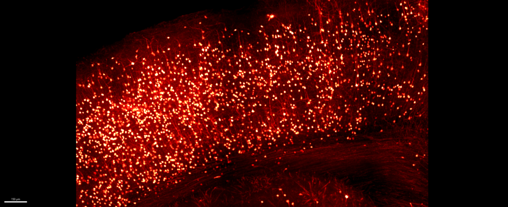 Light my mind by Alessandro VenturinoExperimental details: 1 mm thick mouse brain slice made transparent using CUBIC 2 protocol. System: confocal microscopy, LSM 880 UP, 10X objective. Post-processing: Imaris 9.9.1, cfos-positive neurons = glow.
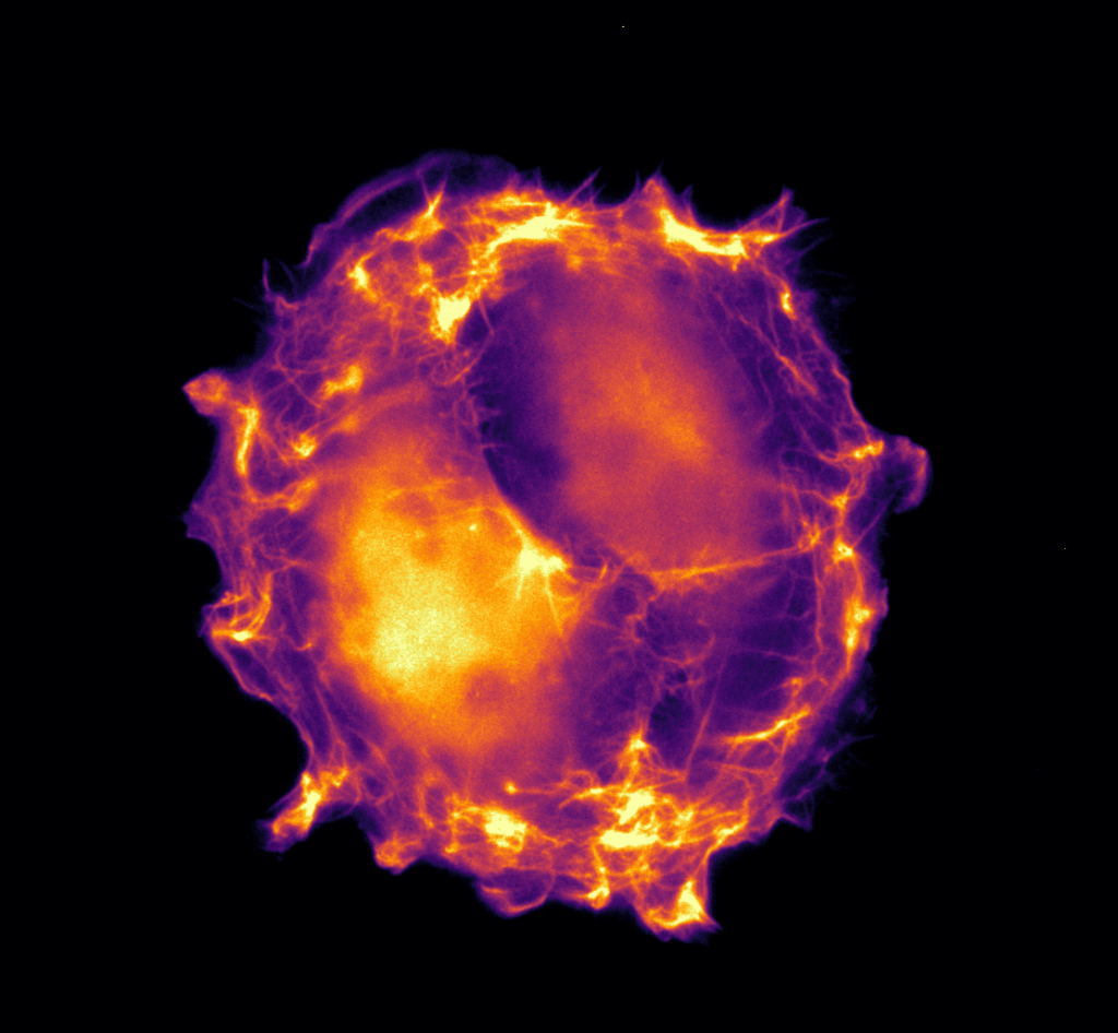 Ying & Yang by Michael RiedlLifeAct-Gfp Endothelial Cell on micropattern acquired on Leica Mica, Brightfield, 63x Magnification, Water Objective
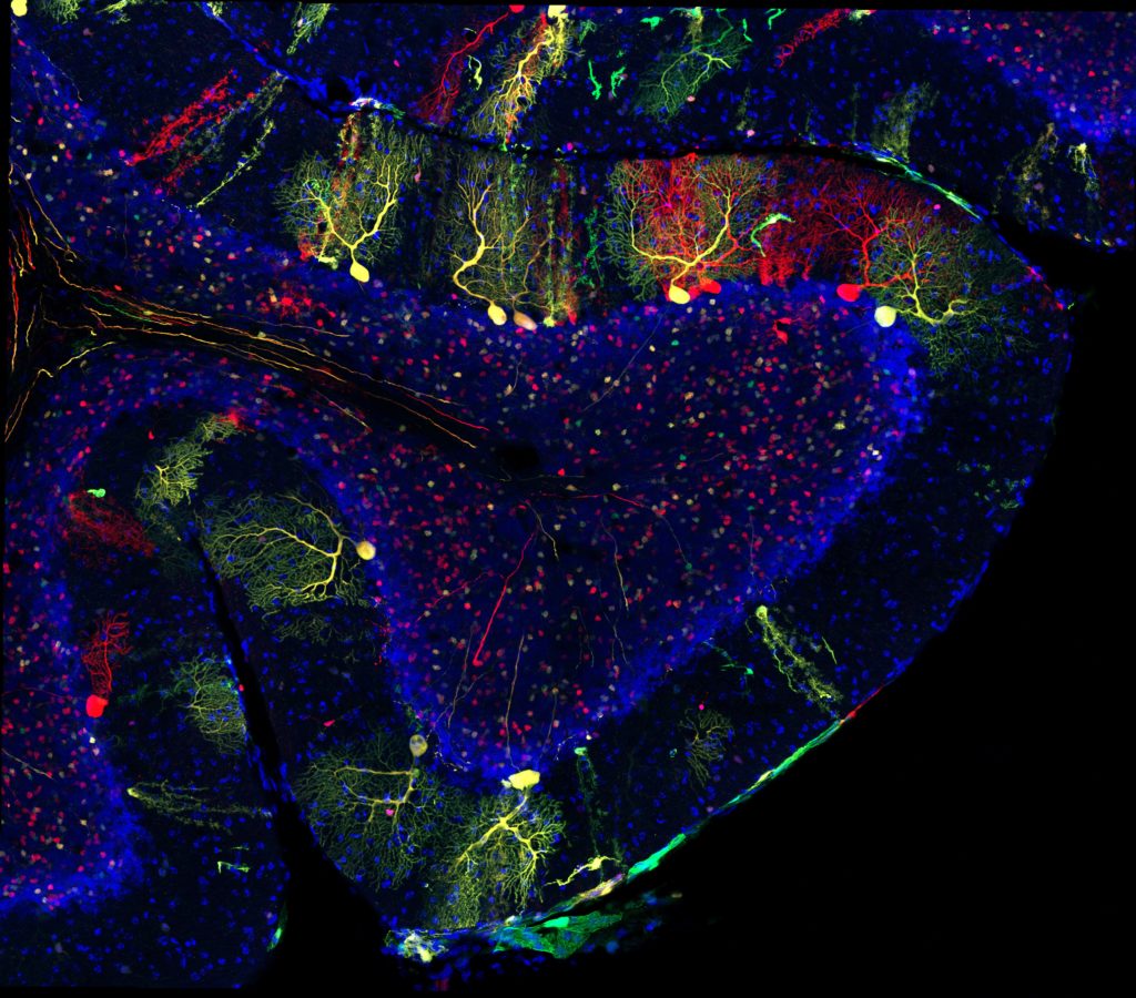 Rainbow Rollercoaster in the Brain by Nicole AmbergMADM-labelled individual red, green and yellow cells in the cerebellum of a 21day old M18-GT/TG Hprt-Cre mouse. The cerebellum is a brain region responsible for coordination and movement of hands and feed, as well as maintenance of posture, balance and equilibrium. The coral-shaped cells are called Purkinje cells. Each Purkinje cell can receive input through almost 100,000 connection sites (so called synapses). They are sending the integrated information from all these synapses through one single axon to the cerebellar nuclei.
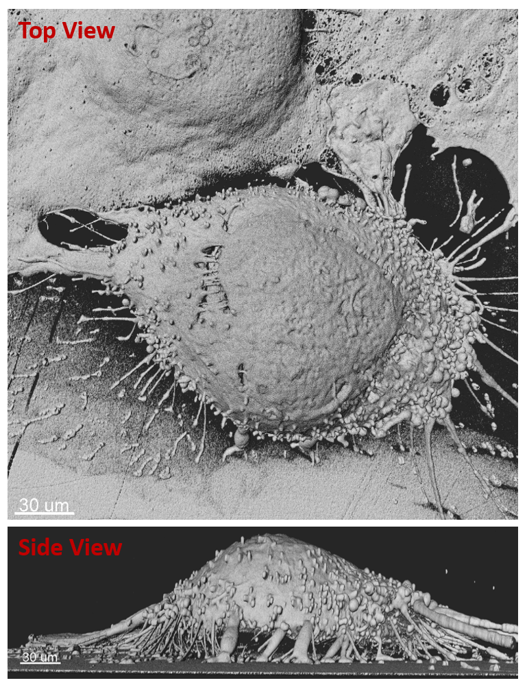 Fasten seat belts, please: the spacecraft will take off soon by Mojtaba TavakoliFour-fold expanded cancer cell getting ready for division. The image was acquired on Andor Dragonfly system
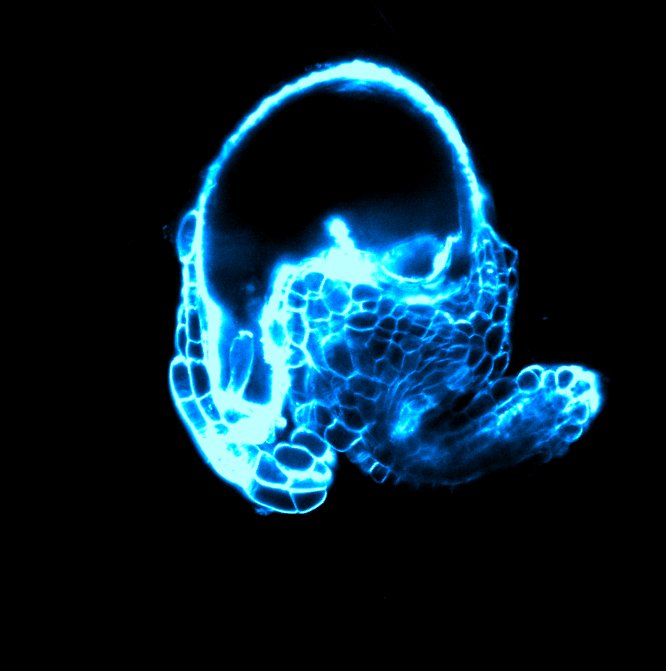 Alien Egg by David BabicAn ovule (immature seed) of the Arabidopsis thaliana plant counterstained with the SR2200 cell wall stain. Acquired on Zeiss LSM 800 confocal microscope.
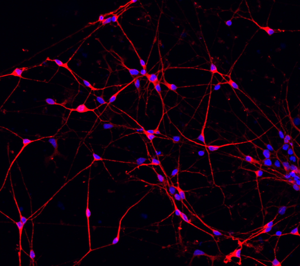 The Brain’s Web by Medina KorkutImage shows neurons derived from human induced pluripotent stem cells that build a neuronal network in 2D culture. Neurons shown in red and nuclei in blue.
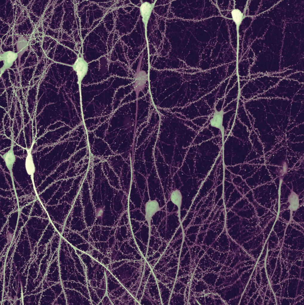 Neuronal deep space by Philipp VelickyThe visible neurons are alive and produce a fluorescence protein. Imaged with the Leica Multiphoton DIVE microscope of the IOF in a mouse hippocampal slice culture. Die sichtbaren Neuronen sind lebendig und produzieren ein fluoreszierendes Protein. Aufgenommen wurde das Bild mit dem Leica Muliphoton DIVE Mikroskop der IOF in einer Schnittkultur eines Maus-Hippocampus.
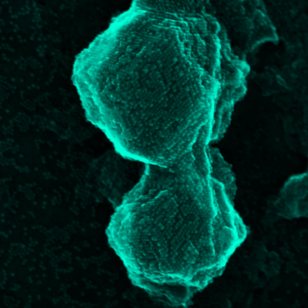 SUPERCRYSTALS! by Seungho LeeSelf-assembled core-shell nanocrystals. Image acquired on JEOL 2800 TEM.
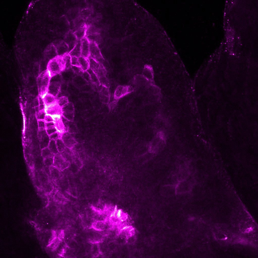 Heart Veins by Caterina GianniniImmunostaining of Arabidopsis thaliana leaf primordia imaged with Zeiss LSM800 confocal system. The image shows the stained membrane protein (magenta) in the vasculature pattern of the new leaf.
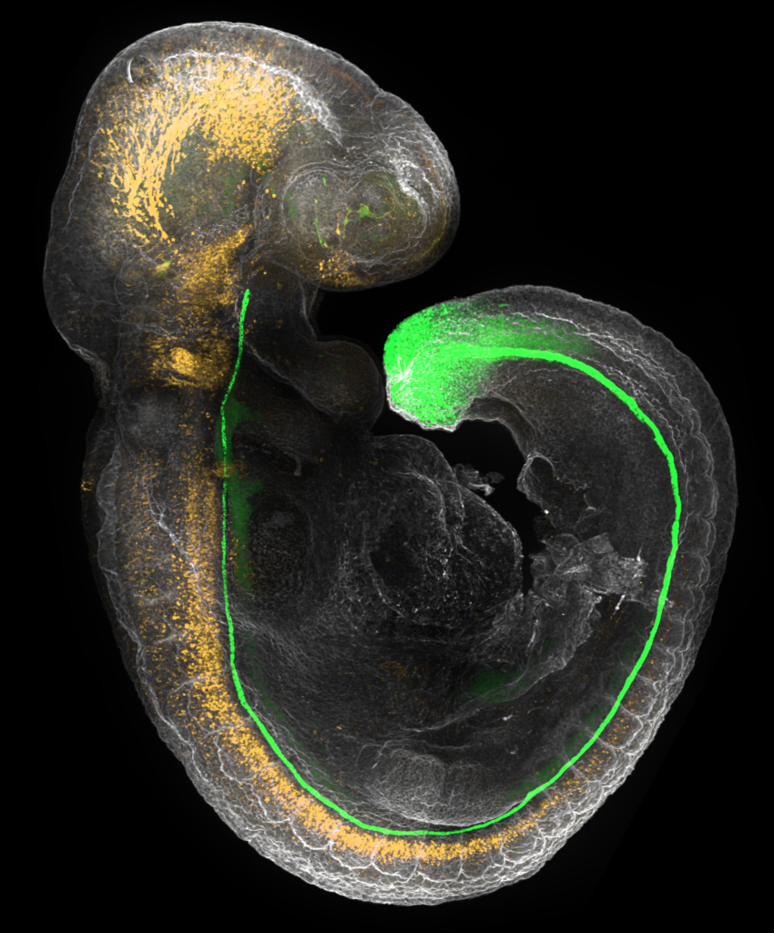 Mouse embryo by Kasumi Kishi9 day old mouse embryo stained with fluorescent antibodies. Neurons are shown in orange and an organ called the notochord in green.
Scroll Up
 Light my mind by Alessandro VenturinoExperimental details: 1 mm thick mouse brain slice made transparent using CUBIC 2 protocol. System: confocal microscopy, LSM 880 UP, 10X objective. Post-processing: Imaris 9.9.1, cfos-positive neurons = glow.
 Ying & Yang by Michael RiedlLifeAct-Gfp Endothelial Cell on micropattern acquired on Leica Mica, Brightfield, 63x Magnification, Water Objective
 Rainbow Rollercoaster in the Brain by Nicole AmbergMADM-labelled individual red, green and yellow cells in the cerebellum of a 21day old M18-GT/TG Hprt-Cre mouse. The cerebellum is a brain region responsible for coordination and movement of hands and feed, as well as maintenance of posture, balance and equilibrium. The coral-shaped cells are called Purkinje cells. Each Purkinje cell can receive input through almost 100,000 connection sites (so called synapses). They are sending the integrated information from all these synapses through one single axon to the cerebellar nuclei.
 Fasten seat belts, please: the spacecraft will take off soon by Mojtaba TavakoliFour-fold expanded cancer cell getting ready for division. The image was acquired on Andor Dragonfly system
 Alien Egg by David BabicAn ovule (immature seed) of the Arabidopsis thaliana plant counterstained with the SR2200 cell wall stain. Acquired on Zeiss LSM 800 confocal microscope.
 The Brain’s Web by Medina KorkutImage shows neurons derived from human induced pluripotent stem cells that build a neuronal network in 2D culture. Neurons shown in red and nuclei in blue.
 Neuronal deep space by Philipp VelickyThe visible neurons are alive and produce a fluorescence protein. Imaged with the Leica Multiphoton DIVE microscope of the IOF in a mouse hippocampal slice culture. Die sichtbaren Neuronen sind lebendig und produzieren ein fluoreszierendes Protein. Aufgenommen wurde das Bild mit dem Leica Muliphoton DIVE Mikroskop der IOF in einer Schnittkultur eines Maus-Hippocampus.
 SUPERCRYSTALS! by Seungho LeeSelf-assembled core-shell nanocrystals. Image acquired on JEOL 2800 TEM.
 Heart Veins by Caterina GianniniImmunostaining of Arabidopsis thaliana leaf primordia imaged with Zeiss LSM800 confocal system. The image shows the stained membrane protein (magenta) in the vasculature pattern of the new leaf.
 Mouse embryo by Kasumi Kishi9 day old mouse embryo stained with fluorescent antibodies. Neurons are shown in orange and an organ called the notochord in green.