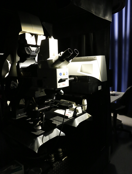
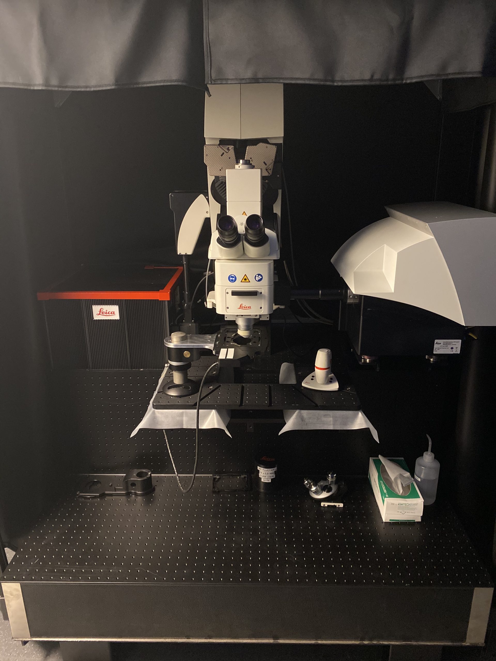
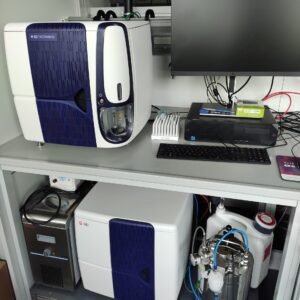
The Leica TCS SP8 DIVE CS is a unique microscope due to its large working distance and ability to handle a wide variety of multi-photon imaging applications, such as deep live imaging, capture calcium dynamics (resonance scanner and fast stage), cleared tissue imaging (special objectives), and complex multi-color acquisitions, such as (b)rainbow, due to the option to dynamically tune the detection windows. In addition, the system allows for life-time imaging in MP, and time-gating of undesired signal (post-processing).
Description
The Leica TCS SP8 DIVE CS is a unique microscope due to its large working distance and ability to handle a wide variety of multi-photon imaging applications, such as deep live imaging, capture calcium dynamics (resonance scanner and fast stage), cleared tissue imaging (special objectives), and complex multi-color acquisitions, such as (b)rainbow, due to the option to dynamically tune the detection windows. In addition, the system allows for life-time imaging in MP, and time-gating of undesired signal (post-processing).
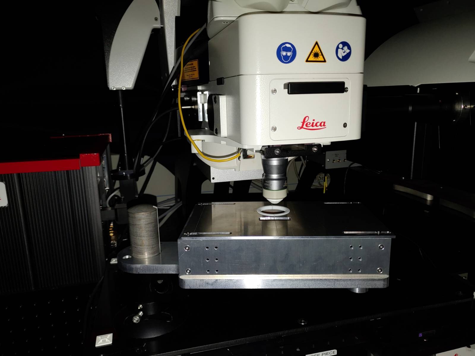
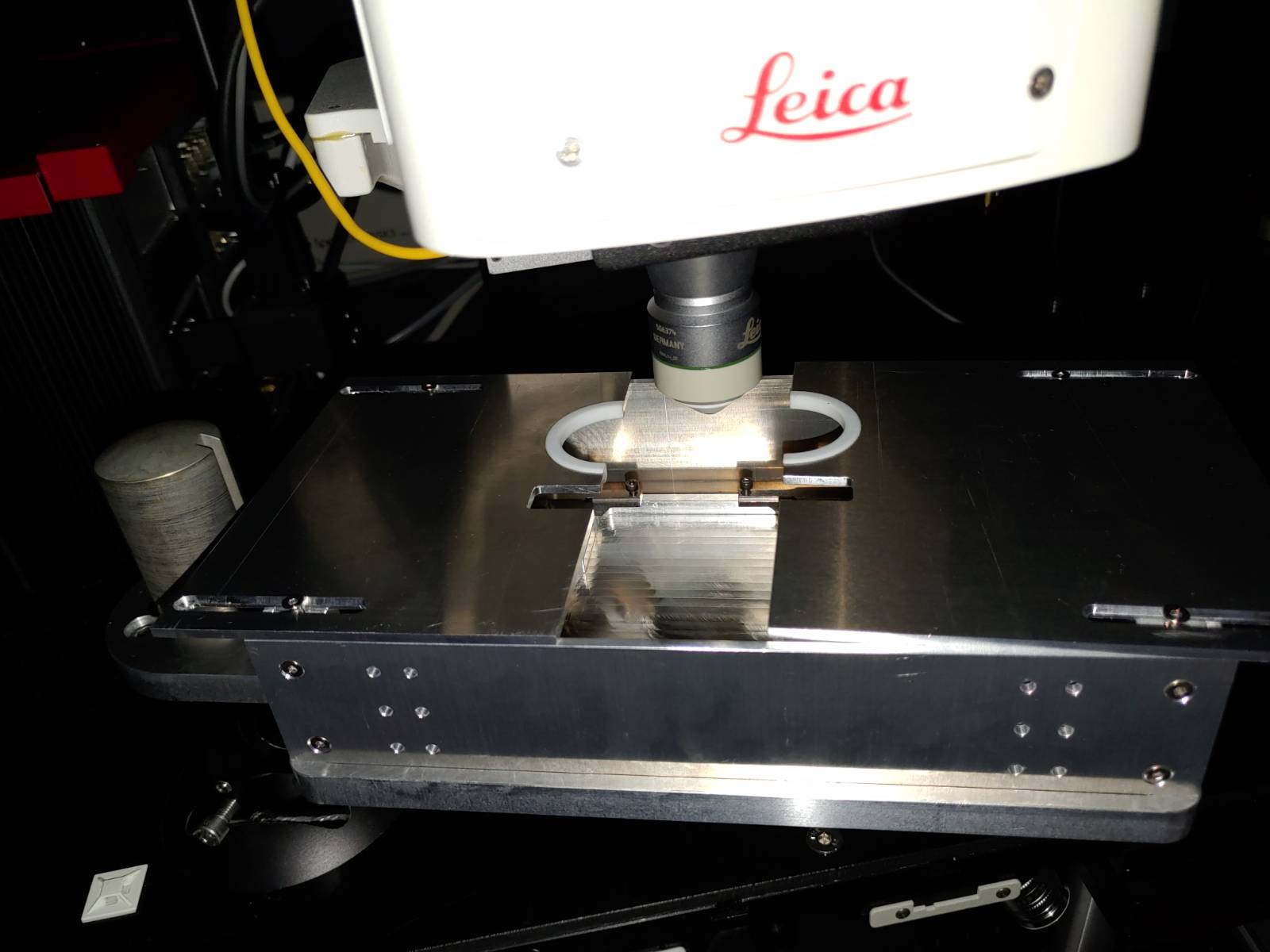
We have designed a new stage with Siegert group to provide Faradey cage for EEG and cranial window recording. (see product gallery)
Location: room i04.U1.018
Imaging modalities/features:
- Multi-photon (680-1300 nm + 1045) – external 4-tune scan head
- Confocal (405nm, 488nm, 552nm, 638nm) – internal scan head
- Second Harmonics Generation (and Third Harmonics Generation)
- Life-time imaging (FALCON, time gated acquisition )
- Real-time deconvolution (Lightning)
- Spectral unmixing
- Excitation Λ / emission λ wavelength scans (in multi-photon)
Specification:
- Leica DMi8 CS fixed stage upright microscope for larger specimen imaging, with cascaded optical table
- TCS SP8 FALCON, with resonance scanner (16000 Hz – bidirectional)
- Scientifica Motorised Moveable Base Plate (MMBP) Scanning stage (step-sice 0.1 μm, range 50mm, max load: 30 kg)
- Super Z Galvo Stage Type H (Maximal travel range in z: 1500 μm Minimal step size: 20 nm)
- Trigger box
Light sources:
- Cool LED fluorescence lamp (visual inspection)
- VIS-Laser: 405nm (50 mW), 488nm (20mW), 552nm (20mW), 638nm (30mW) diodes
- Multi-photon: IR Laser Spectra Physics InSight DeepSee (680-1300 nm + Dual 1045)
Filter for visual examination:
- Triple band-pass → CFP/YFP/RFP: BP 436/20, BP 503/21, BP 575/25 Emission: BP 470/25, BP 535/30, BP 630/70
Objectives:
- Default: Objective turrent (27mm):
- HC PL Fluotar 10x/0.30, WD 2.2mm – no immersion (VIS only)
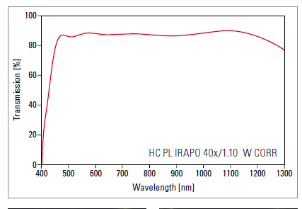
- HC PL APO 40x/1.10 W CORR CS2, WD 0.65mm – water immersion, Correction collar for use with 0.14 – 0.18 mm coverglass. (470-1200 nm)
- HC PL APO 63x/1.40 OIL CS Plan, WD 0.14mm
- HC L Fluotar 16x / 0.60 MI (# 155065), WD 2.5 mm – ne 1.33-1.52 (water, silicon- or glycerol immersion – (Correction collar for use with 0.0 – 0.17 mm coverglass) + BABB cleared samples
- Single objective: HC FUOTAR L 25x / 0.95 W (# 15506374), WD=2.5 mm, wide angle (41°)
- Empty
- Additional high-end rental objectives:
- Default objective – Single objective holder (32mm):
- HC IRAPO L 25x/1.00 W mot IRAPO, WD 6.2mm – water, motoCORR (470-1200 nm) – €€€ extremely high-value objective
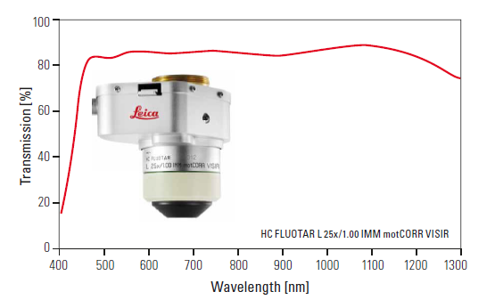
- HC IRAPO L 25x/1.00 W mot IRAPO, WD 6.2mm – water, motoCORR (470-1200 nm) – €€€ extremely high-value objective
- Single objective holder (32mm):
- HC IRAPO L 16x/0.8 W mot VISIR, WD 8.15mm – water, motoCORR (470-1200 nm) – €€€ extremely high-value objective
- Default objective – Single objective holder (32mm):
Internal scan head detectors: confocal- mode
- PMT
- HyD SP GaAsP Detector
- HyD SP GaAsP Detector
External (4-tune) NDD detectors: multi-photon mode
- PMT
- HyD2 SMD: specially selected for Falcon FLIM
- HyD2 SMD: specially selected for Falcon FLIM
- PMT
Modules: Computer (HPZ840 Workstation – Win 10 professional – 64 bit): System manual and additional material is available on SeaFile (login required). Contact: iof@ist.ac.at Request a training: training request form
Additional information
| Building | i04 |
|---|---|
| Camera / Detector | MA-PMT, Hyd |
| Fluorescent Lightsource | Laser: 405±5 nm, Laser: 488±5 nm, Laser: 640±5 nm, Laser: 1045nm MP, Laser: 680-1300nm MP |
| System_Specification | Illumination: Fluorescence, Illumination: Bightfield, Incubation: heating stage, Automated Stage, Z-piezo / galvo Stage |
| Technology / Application | FLIM, FRAP, FRET, FRET-FLIM, Time-lapse recording, Multi-position, In-vivo imaging, Intravital imaging, Ablation: UV/IR-laser based, Photoactivation / -conversion, SHG, Spectral Imaging |
| Custom Tools / Inserts | Cooling stage-insert compatible (Lab) |

